Carotid Body Tumor
Carotid body tumors (CBTs) are rare neoplasms; although they represent about 65% of head and neck paragangliomas (These develop at various body sites including the head, neck, thorax and abdomen). These are located on the side of the neck, where the large carotid artery branches into smaller blood vessels to carry blood into the brain. The cluster of cells around that branching is called the carotid body, or carotid glomus. The tumors that develop there are not life-threatening, but they can grow quickly and press on nearby nerves and blood vessels, causing damage to those structures.
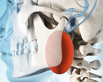
The carotid body, which originates in the neural crest, is important in the body’s acute adaptation to fluctuating concentrations of oxygen, carbon dioxide, and pH. The carotid body protects the organs from hypoxic damage by releasing neurotransmitters that increase the ventilatory rate when stimulated.These tumours arise from chemoreceptor zone present at the division of carotid artery (supplying arterial blood to face – external carotid artery and brain- internal carotid artery) into external and internal carotid artery as shown in the figure.
3 Signs and Symptoms
Usually presents as a painless neck mass, but larger tumors may affect cranial nerve , usually of the vagus nerve (leading to change or heaviness of voice) and hypoglossal (tilting or deviation of tongue towards the side of tumor) nerve. These are highly vascular tumors means have many small blood vessels around it.Individuals with malignant (cancerous) carotid body tumors may have higher blood pressure.
Types
- Sporadic: The sporadic (does not genetic link) form is the most common type, representing approximately 85% of carotid body tumors.
- Familial: The familial (have genetic link) type (10-50%) is more common in younger patients.
- Hyperplastic:The hyperplastic form is very common in patients living at a high altitude (> 5000 feet above sea level) due to decrease level of oxygen.
About 5% of carotid body tumors are present on both sides of neck and 5-10% can be cancerous also, but these rates are much higher in patients with inherited disease.
Diagnosis
The test performed are ultrasonography with colour Doppler, CT scanning of the head and neck with contrast or CT Angiography is also helpful and typically reveals a hyper vascular tumor located between the external and internal carotid arteries.
MRI imaging may also be considered to be the criterion standard of carotid body tumors, and the tumor has a characteristic salt and pepper appearance on T1-weighted image.
Treatment
Surgical excision is done by taking care of nearby vital structures and utmost care of internal carotid is observed.The process goes as under general anaesthesia, laterally inclined incision along the anterior border of the sternomastoid muscle is performed intraoperatively. After clear visualization of the anatomic structures such as the common carotid artery, the internal carotid vein, the cranial nerve, and the accessory nerve, then the common carotid artery and the internal and external carotid veins are sufficiently liberated for a good separation, and then the common carotid artery and the proximal end of the tumor artery are blocked, using blood vessel blocking bands for the convenient control of the blood flow, and finally they are carefully separated along the tumor body so that the blood vessels feeding the tumor could be radically removed. After the removal process, Reconstruction of the common carotid and the internal carotid artery are performed in patients undergoing resection of the internal carotid artery. Patient can be discharged in 1-2 days.


